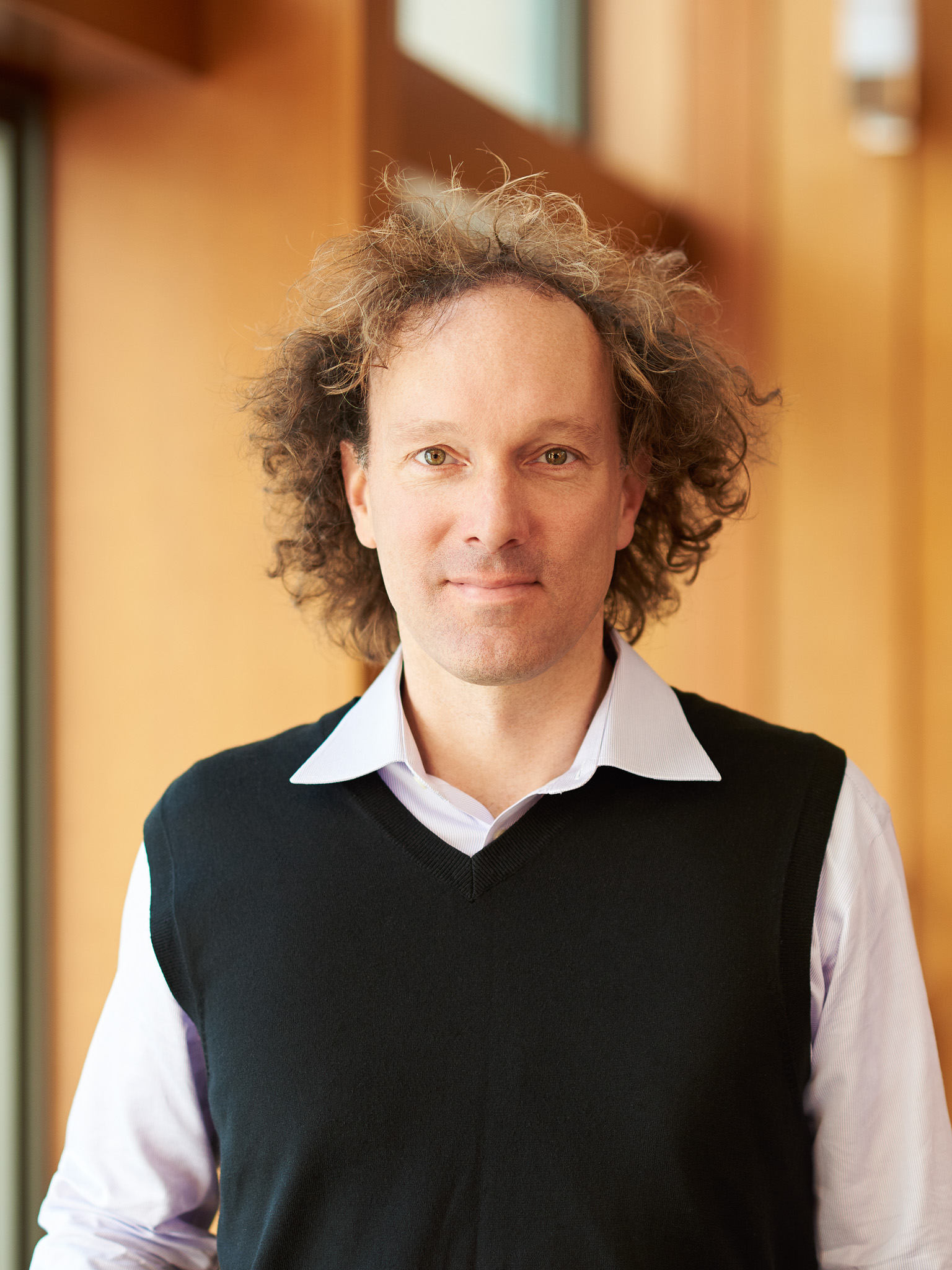
Daniel Fletcher
BioengineeringDaniel Fletcher is the Chatterjee Professor of Engineering Biological Systems and Chair of the Department of Bioengineering. His laboratory develops medical devices and studies biophysical mechanisms of disease, with a particular interest in infectious diseases and global health. He received his BS from Princeton University, his D.Phil from Oxford University as a Rhodes Scholar, and his PhD from Stanford University.
Spark Award Project
Mobile Phone-Based Eye Care
Routine eye exams are required for effective diagnosis and management of diabetic retinopathy, glaucoma, and other major causes of blindness. Despite coverage of exam costs by insurance and Medicare, many people forgo necessary screening. A low-cost handheld retinal imaging tool could increase access to routine eye exams by making high-quality retinal imaging available from primary care physicians and other healthcare professionals. Together with collaborators, we have developed a mobile phone-based ophthalmoscope capable of capturing wide-field retinal images for rapid screening of retinal diseases.
Daniel Fletcher’s Story
Your cell phone can already find your car and tell you what song the restaurant is playing. How about an app to screen for eye disease?
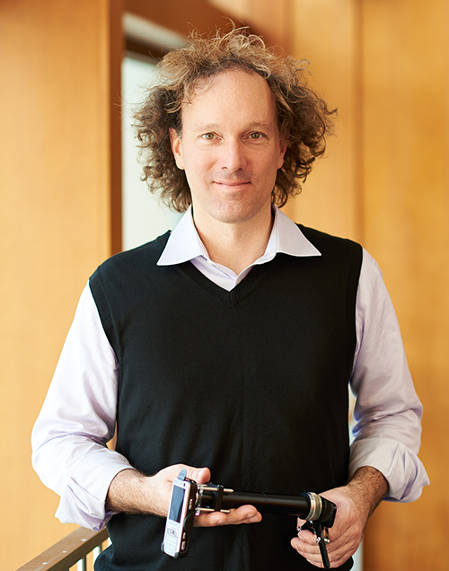
By coupling the sophisticated imaging capabilities of smart phone cameras with lenses and software for examining the retina, Daniel Fletcher and his students have developed a hand-held, user-friendly version of the optometrist’s ophthalmoscope and are teaming up with clinical collaborators to detect retinal disease caused by diabetes.
“The advanced technology built into a cell phone is allowing us to scale down what would normally be much larger medical devices,” says Fletcher, Professor of Bioengineering. “The key is the phone, which we modify with hardware and software to make it a tool for physicians, and eventually consumers.”
The RetinaScope could allow a primary care physician to easily distinguish a healthy retina from one in the early stages of retinopathy, a diabetes-related malady that can lead to blindness. Currently, the exam is done only by eye specialists, who use table-top ophthalmoscopes to examine the delicate branching of the retina’s blood vessels. If retinopathy has developed, blood vessels can hemorrhage, killing retinal cells. But if it is detected early, it is treatable.
“This device can cut out a step,” Fletcher says, “so your primary care doctor can screen for retinal abnormalities during a regular checkup. If something indicates there is a problem, then you can be referred to an ophthalmologist.”
Clinical results from pilot studies at medical centers of the University of Michigan and Washington University in St. Louis are showing the practicality and reliability of the RetinaScope as a screening tool for retinal diseases. In a way, that may be the easy part.
“One big challenge, as with any medical technology, will be developing a business model that will work with insurance companies,” Fletcher says. “We’ve made a lot of progress on the technical side, but moving into the commercial world, getting it into broader use — that’s a different issue.”
For that he has support from the Bakar Fellows Program.
“I think that’s exactly what the Bakar funding can do —- help us refine the technology and clinical use case so that we can come up with a viable business plan that sustainably increases access to quality eye care.”
This isn’t his lab’s first foray into a smart phone-enabled medical devices. In 2009, Fletcher’s students developed a mobile phone-based otoscope, an instrument used to detect ear infections. They formed a startup company, CellScope Inc, and the instrument is now on the market.
“As a kid, I had a lot of ear infections. My parents would take me to the doctor, who would stick a little magnifier into your ear and decide whether you have something that needs to be treated or not — just by looking. Why not use the camera of your phone for that purpose, in the comfort of your own home?”
Overall, Fletcher aims to make medical diagnosis and care less expensive and more accessible, and not just for the developed world.
His research team is currently working on a project to expand treatment for river blindness in West Africa using a portable mobile-phone based microscope he calls the LoaScope. Its software can examine a pinprick of blood to determine if a drug for the disease can be safely given to patients.
“Just as they have done for communication, mobile phones can now do for healthcare, making high quality health disease diagnosis and screening possible even hundreds of miles from a medical facility,” Fletcher says.
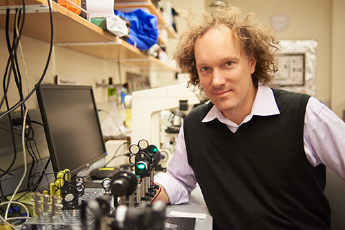
Much of the momentum to build mobile phone-based medical devices comes from UC Berkeley students, Fletcher says. The original mobile phone microscope idea was first demonstrated in 2006 in an optics class he teaches. Students from the class were involved in building the first device, and students at all levels continue to be key contributors.
“Building these devices takes a range of skills. The students are the connectors. Sometimes it’s a friend of a friend who adds the expertise that’s needed.
“The cell phone’s technology is so sophisticated, it’s really amazing what they are capable of compared to even five years ago. When we buy a phone, we are paying for all that technology. We’re buying convenience, but it can also be used to improve people’s lives, even save their lives. It’s a virtuous circle.”