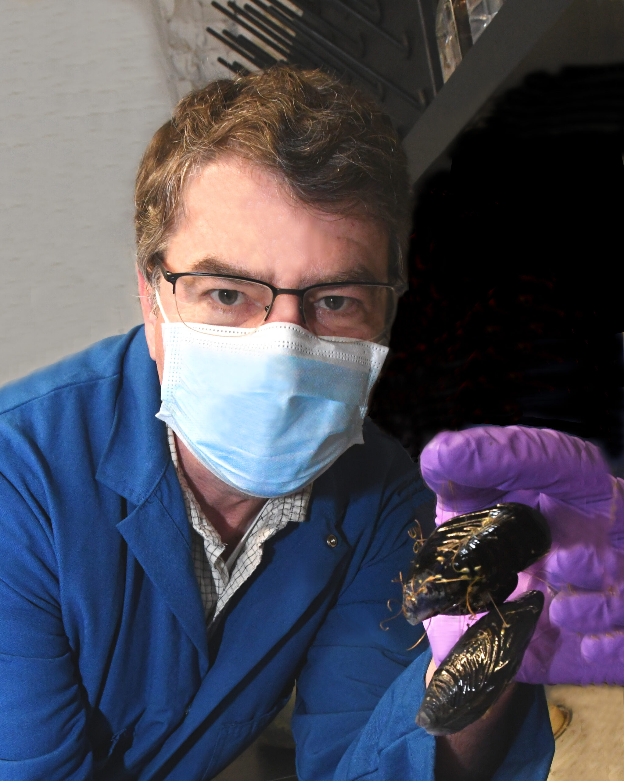
Phillip Messersmith
BioengineeringMaterials Science and EngineeringPhillip Messersmith is a Professor of Bioengineering and Materials Science & Engineering. He earned his BS from the University of Illinois at Urbana and his MS from Clemson University. He earned his PhD in Materials Science & Engineering at the University of Illinois at Urbana.
Spark Award Project
The Messersmith lab focuses on applying structure-property relationships in biological materials to healthcare technology. His current projects include molecular mechanochemical studies of biological wet adhesives, the design of biomimetic wet adhesive polymers and polymer composites, the control of biointerfacial phenomena in nanosystems and in antifouling surface coatings, and the development of novel biomaterials for regenerative medicine.
Phillip Messersmith’s Story
With no known cause and no cure, inflammatory bowel disease (IBD) disrupts the lives of more than six million people. Anti-inflammatory drugs treat the damaged tissue lining the intestine, but the condition flares again about half of the time. Up to a third of people with IBD have surgery to remove part of the GI tract, and many need continued treatment that can triple health care costs.
A combination of two insights – one inspired by mussels’ ability to adhere to rocks – has led Phillip Messersmith to a novel strategy and possibly a potential treatment for IBD.
Messersmith, a professor of bioengineering and materials science and engineering, is developing a way to deliver a drug to damaged tissue in the intestines and assure the drug will stick to the damaged sites to promote healing.
He describes the inspiration and the research supported by his Bakar Fellows Program Spark Award to demonstrate the promise of the regenerative strategy.
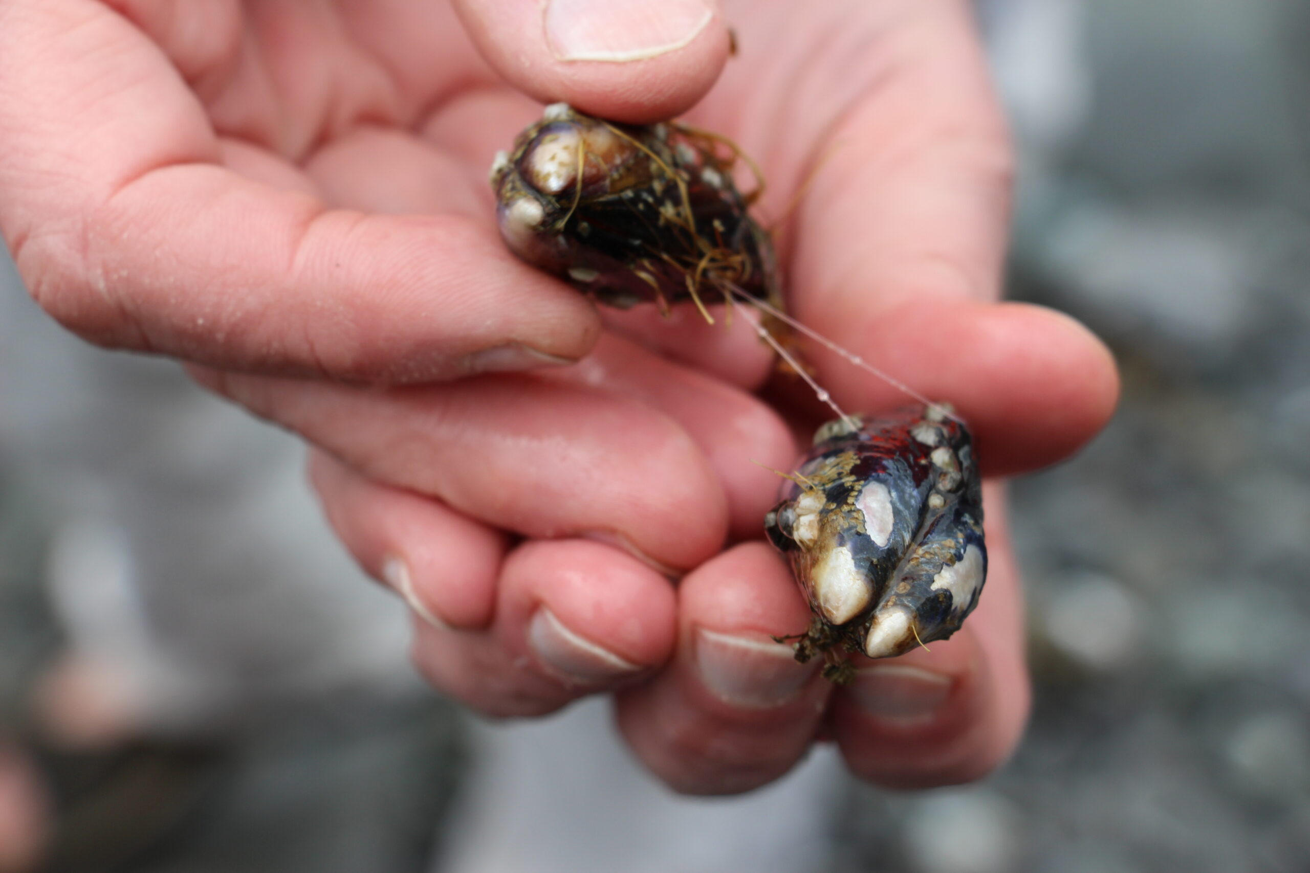
Q) What enables mussels to attach so tightly to rocks and how does your research exploit that ability?
A) Mussels are famous for adhering to pretty much anything, using specialized proteins to glue themselves to rocks along the coast, but also to wood, to whales and other organisms. My lab has studied the adhesion properties of mussel glue proteins for 25 years trying to learn how these proteins work as glues.
A component of that protein, an amino acid called L-dopa, is found in mussels in high concentration. We have spent a lot of time distilling what’s important about L-dopa in relation to adhesion. The materials we develop don’t mimic the mussel protein, but they are inspired by it – they are inspired by nature.
Q) How did your thinking go from mussel adhesion to a possible cure for such an intractable disease?
A) Our work has given us a lot of ideas about how to enhance the binding of drugs to tissues.
We have a project on periodontal disease. In advanced states of the disease, the bone anchoring the teeth to the jaw degrades because of runaway inflammation in this little gap. We are developing a technology that exploits L-dopa to deliver a drug to damaged mucosal tissue in the jaw and enable the drug to bind to those sites and regenerate the tissue.
A colleague of ours works with a strain of mice that are “super healers”, meaning they heal wounds remarkably well. She discovered the molecular mechanism underlying this trait and together we developed a drug to reproduce this in other animals.
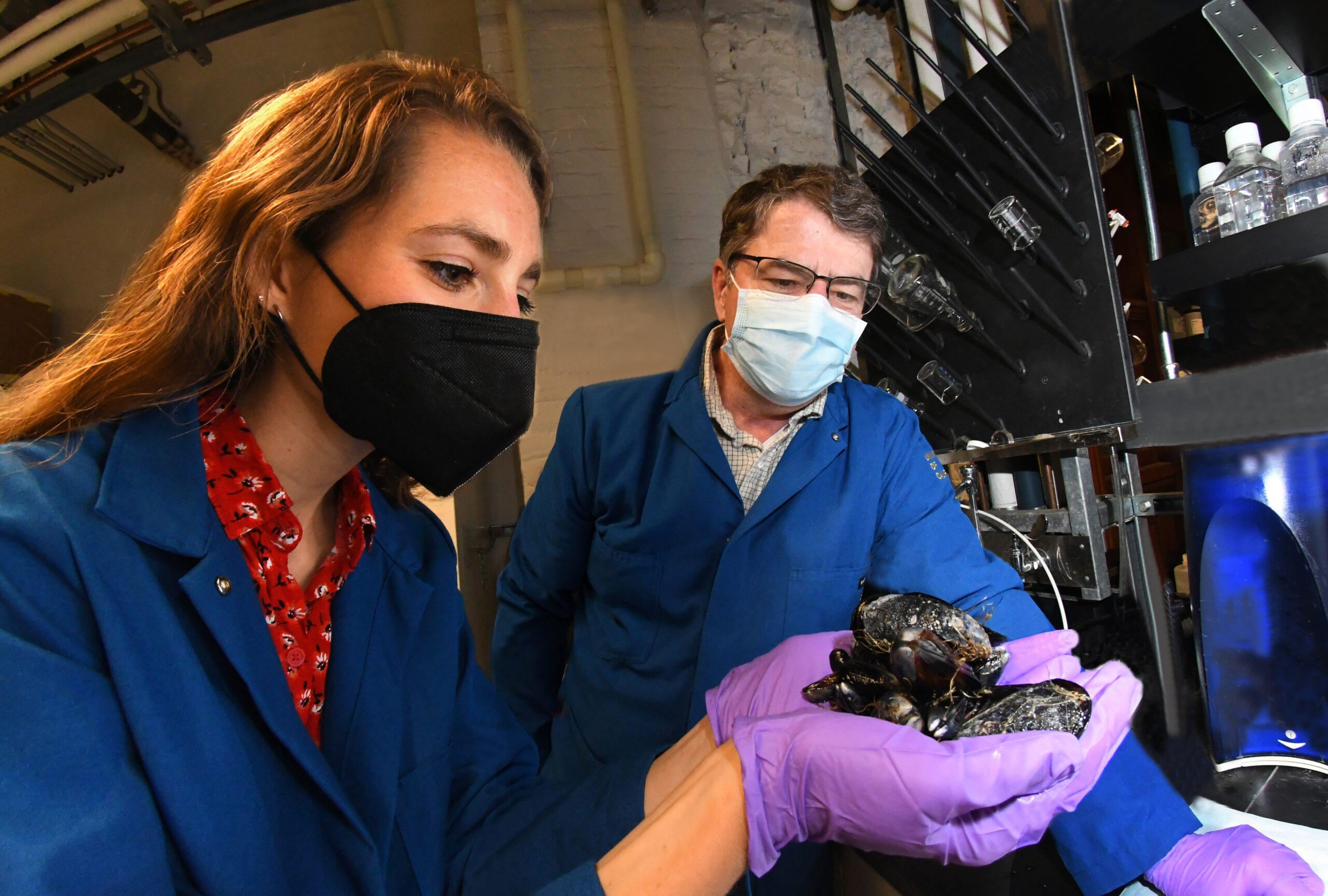
My graduate student, Kelsey DeFrates, proposed to adapt our system to deliver the drug to the lining of the intestine and regenerate IBD lesions. She was present with me when we made the presentation to the Bakar committee.
The intestine is a pretty long organ. How is the drug delivered, and how does it get to the right site in the intestine?
Yes, we want the drug to selectively bind to the lesions. In this tissue there is a breakdown of the mucosal barrier that exposes the underlying basement membrane, which has a certain electrostatic charge that we can exploit to stick the drug to the lesion.
That is amazingly serendipitous. It’s as if the lesions are broadcasting their diseased state and you are able to home in on it.
Yes, it should allow us to get the drug to the right target. That’s the idea.
How will you know that the drug has been delivered to the sites where it is needed?
We use fluorescence – a widely used technology in which a molecule emits light. We can tag a particle or the L-dopa modified drug with a fluorophore and administer it orally to the animal. After waiting a certain period of time, we use a very sensitive box with a camera to image where the fluorescence occurs. That is where the drug has adhered to the intestinal lining.
This technology is so sensitive that you can even detect the fluorescence in the animal in subsurface tissues. So this part of the project is to confirm that we can selectively target sites in the lining of the intestine. Then in collaboration with our colleagues we will determine if the tissue actually regenerates.
So there are two magical ingredients, so to speak, that are familiar to us – the experimental drug and our mussel-inspired drug delivery system. We have not put them together before in this way. I think that during the three-year Bakar Fellows project we can demonstrate the effectiveness of the tissue regeneration technology in a small animal model. I hope that after some years it can make its way into clinical practice.